- Reaction score
- 6,244
cut and paste
Many of the colour lithographs below were created for US surgeon Joseph Pancoast’s (November 23, 1805 – March 6, 1882) 1844 book A Treatise on Operative Surgery. The blurb tells us: “A treatise on operative surgery comprising a description of the various processes of the art, including all the new operations; exhibiting the state of surgical science in is present advanced condition; with eighty plates containing four-hundred and eighty-six separate illustrations.” These images are for the book’s second edition of 1846, for which they were “enlarged”. Other images can be found in the 1848 work Précis iconographique de médecine opératoire et d’anatomie chirurgicale by Claude Bernard (1813-1878). They are captivating and unsettling.
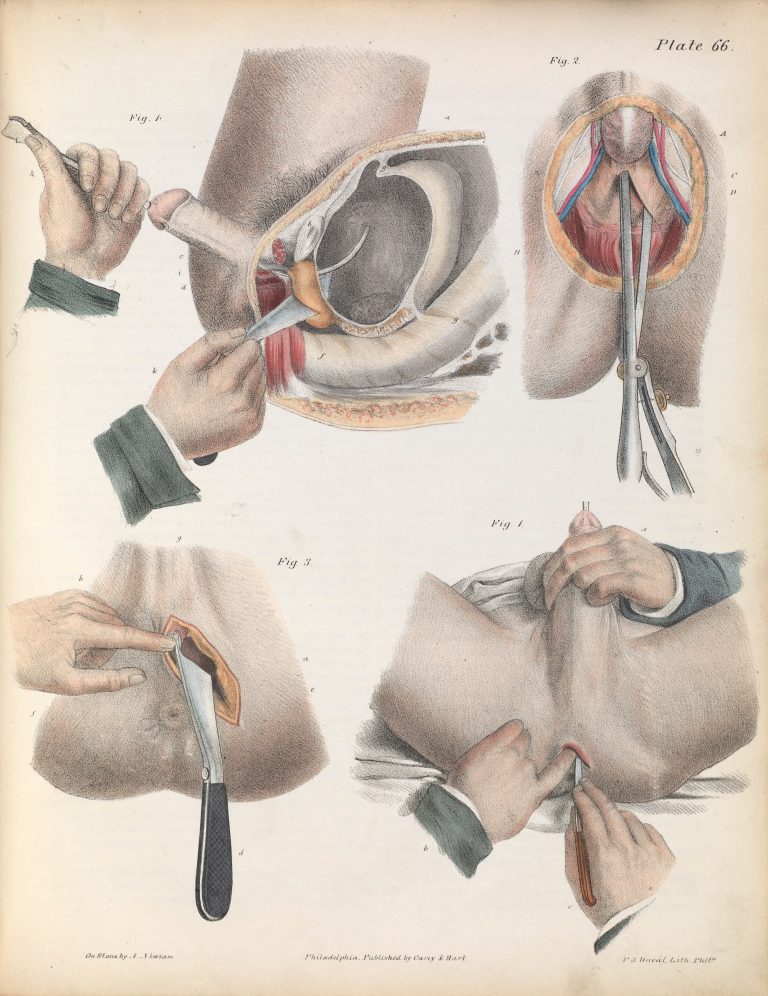
Plate LXVI. Surgical technique for lithotomy.
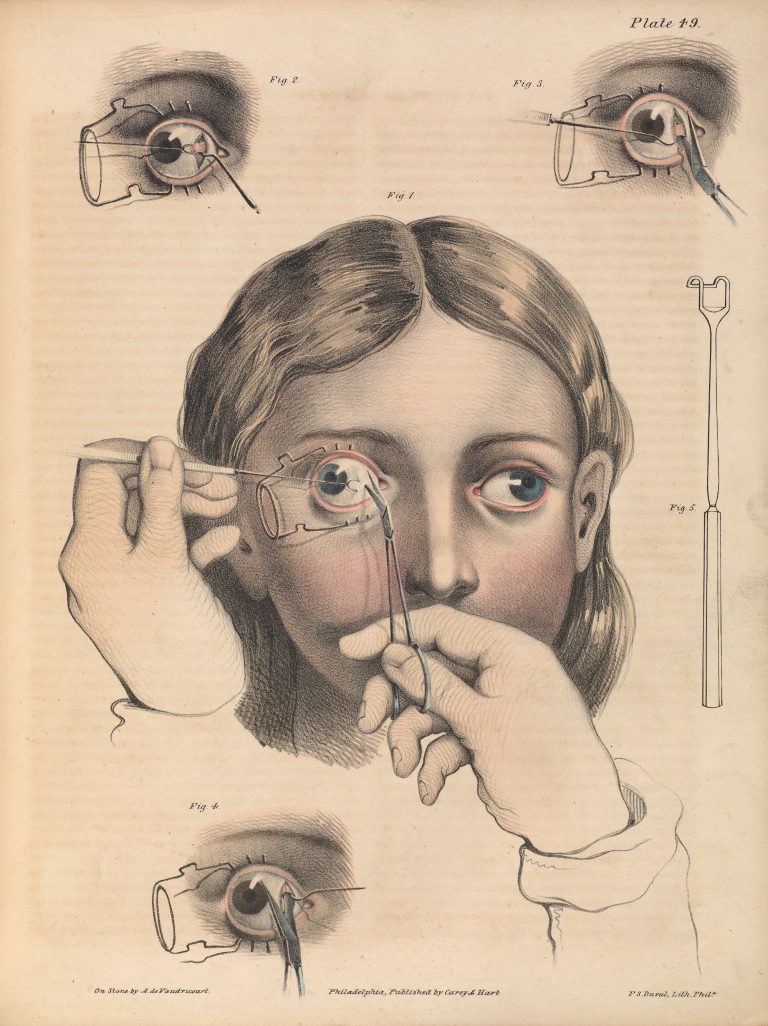
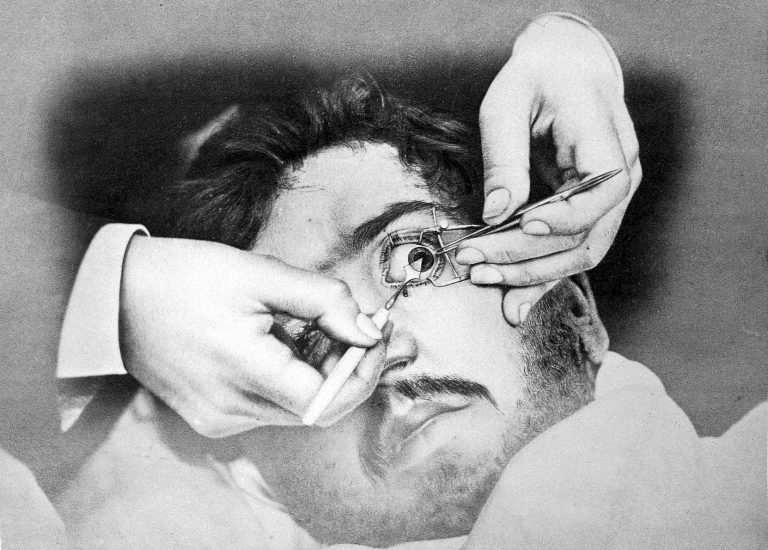
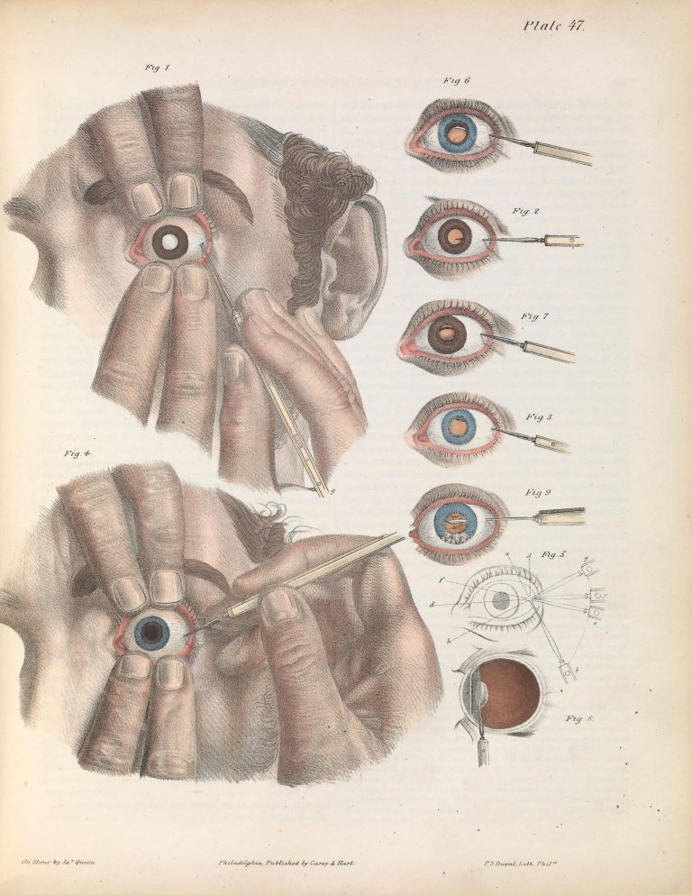
Plate XLIX. Surgery to correct strabismus, involving the division of the internal rectus of the right eye. Strabismus is the misalignment of the eyes.
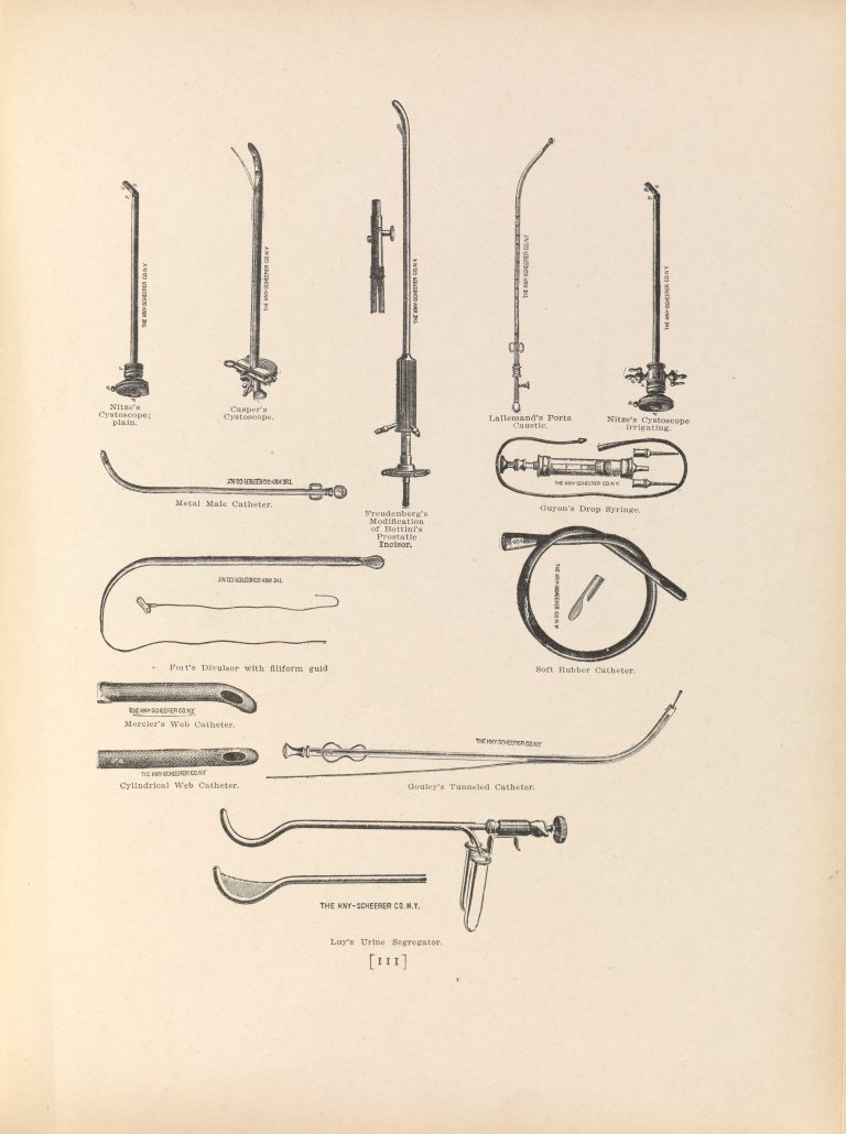
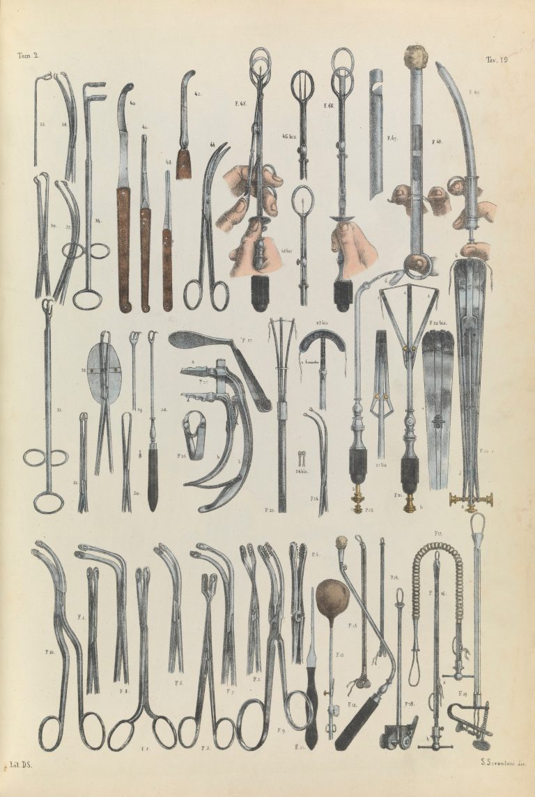
Plate XXXIV. Surgical instruments used for external urethrotomy in prostatectomy (removal of part of the prostate gland).
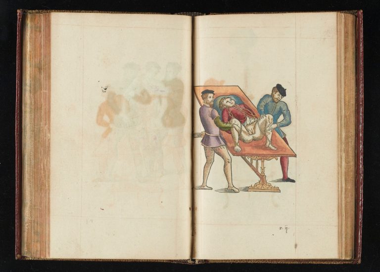
Removing the placenta and umbilical cord after birth. Abbildungen aus dem Gesammtgebiete der theoretisch-praktischen Geburtshülfe, nebst beschreibender Erklärung derselben / Nach dem Französischen des Maygrier bearbeitet und mit Anmerkungen versehen von Eduard Casp. Jac. von Siebold – 1829
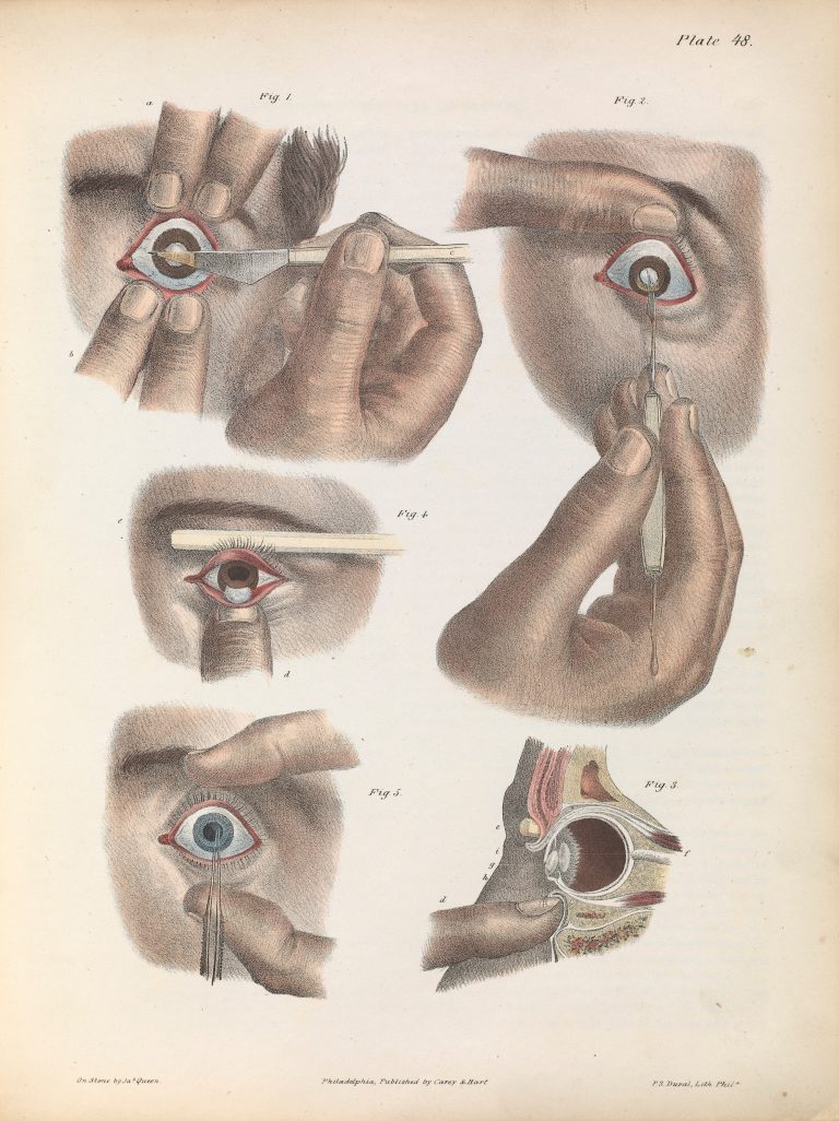
Plate XLVIII. Illustration of surgery on the eye for the removal of a cataract. Operation by extraction – inferior section of the cornea.
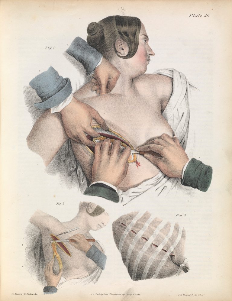
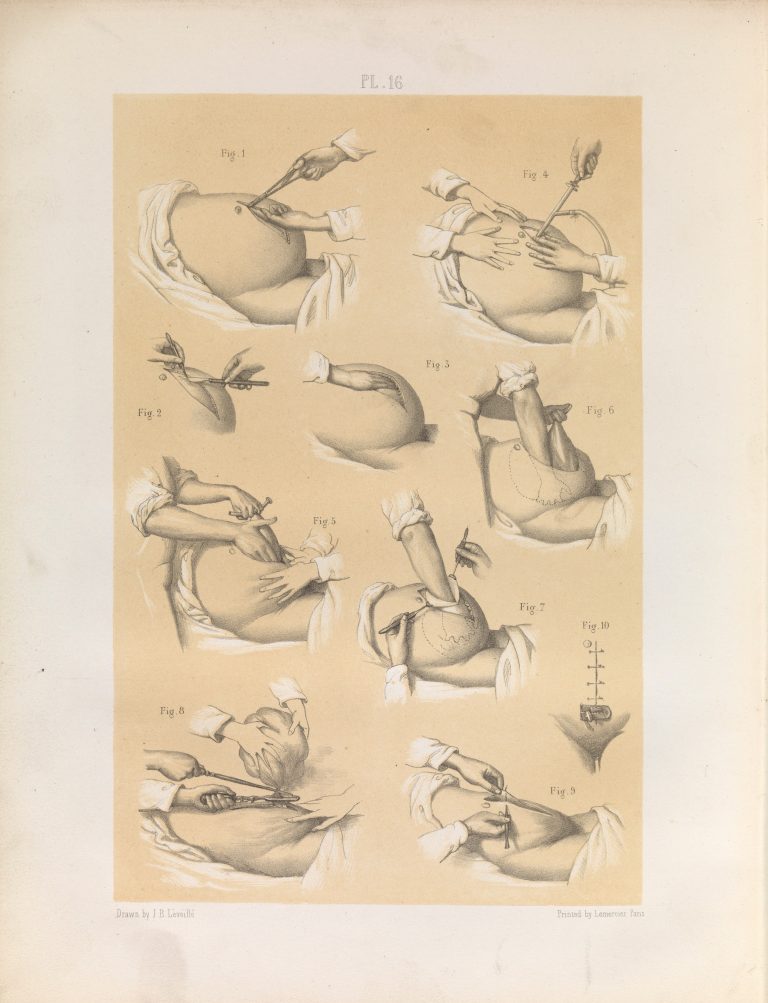
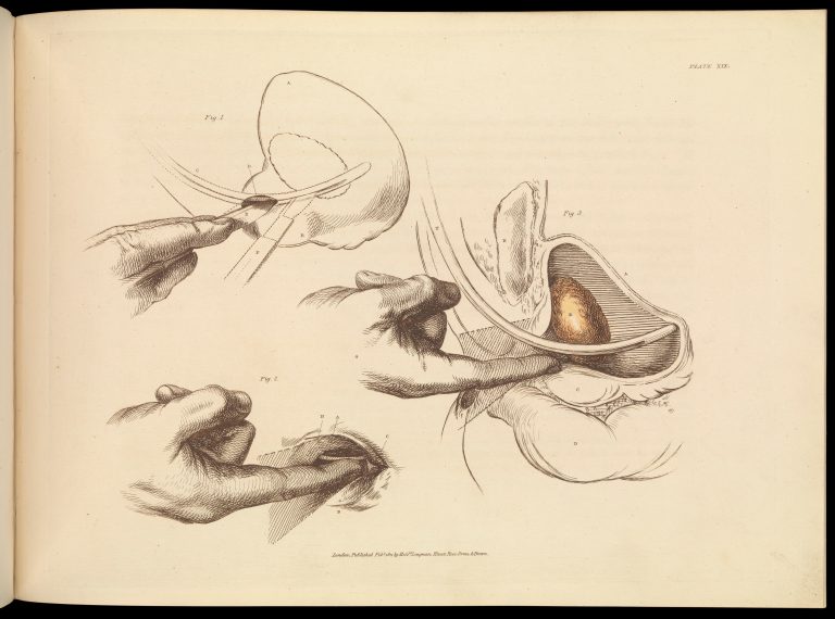
Plate LVI. Surgery for the removal of the mammary gland. 19th Century. Iconografia d’anatomia chirurgica e di medicina operatoria / [Jean Baptiste Marc Bourgery]
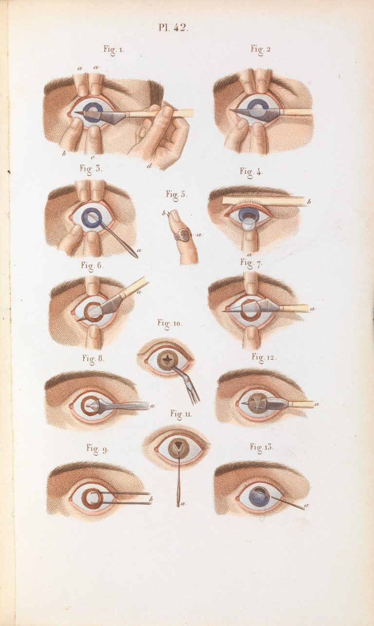
Plate 42, Techniques for the removal of cataracts.
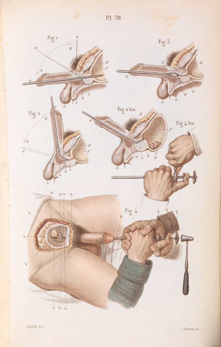
Plate 70, Surgical techniques for lithotripsy (the removal of bladder and kidney stones). Précis iconographique de médecine opératoire et d’anatomie chirurgicale by Claude Bernard (1848).
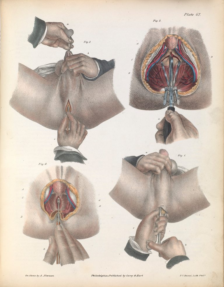
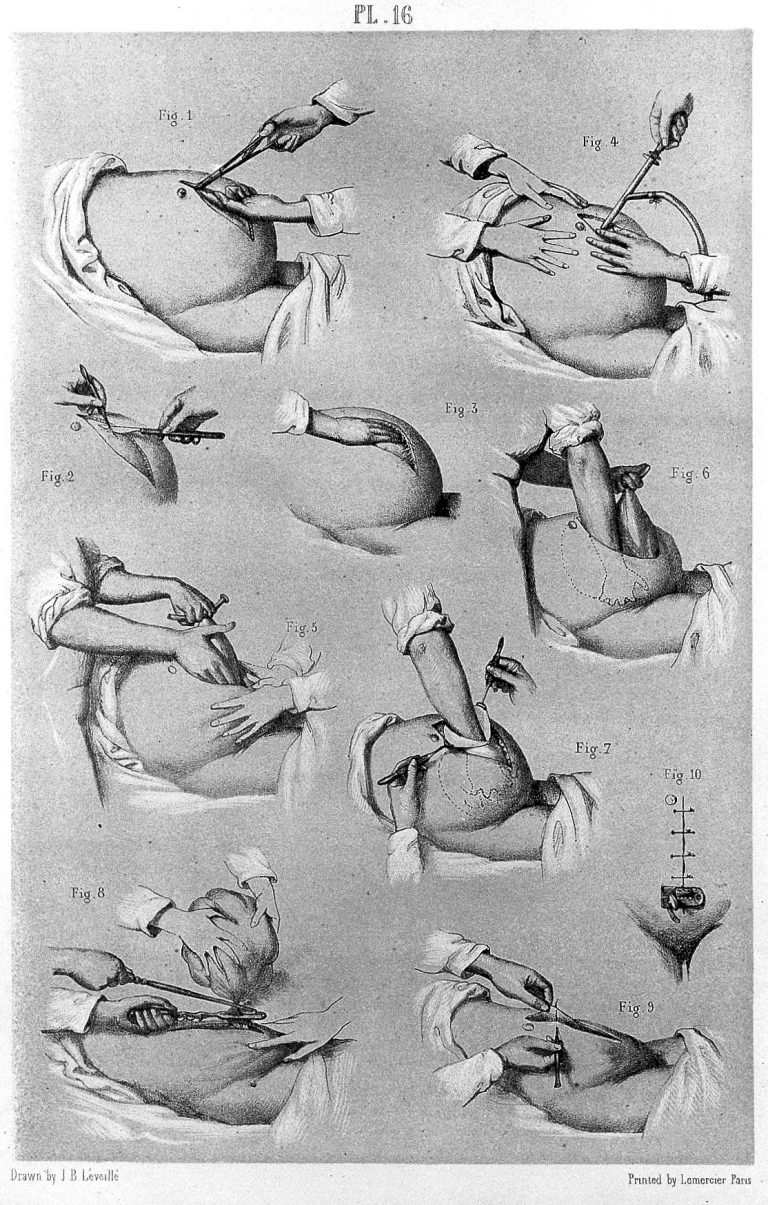
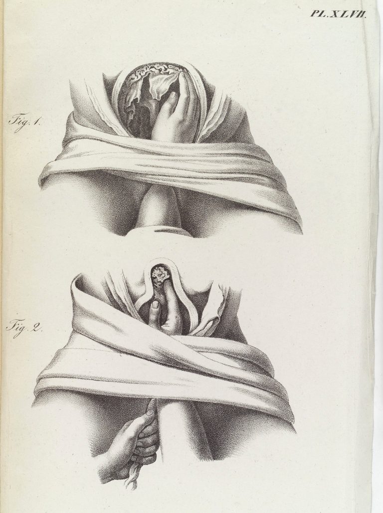
Plate LXVII. Surgical technique for lithotomy (the removal of a bladder stone). Bilateral and vesico-rectal operation.
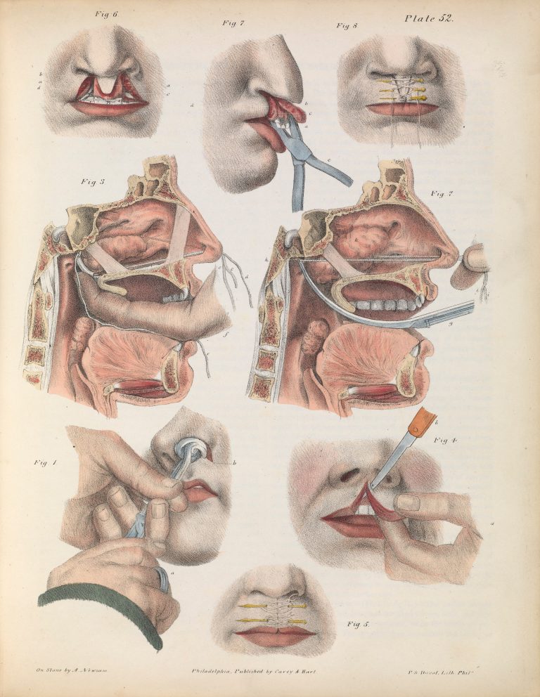
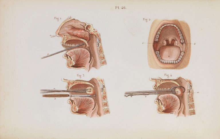
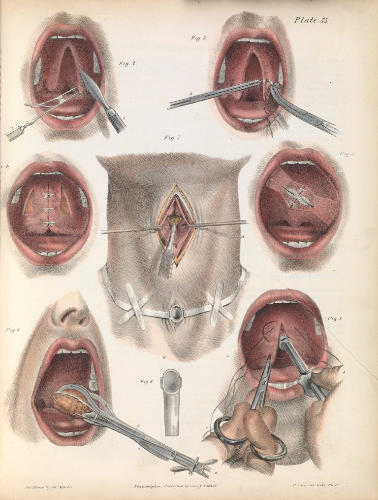
Fig. 1. Removal with the forceps by torsion and traction. Fig. 2, 3. Removal by ligature. Fig. 4,5. Simple hare-lip. Fig. 6, 7, 8. ‘Double hare-lip’ and ‘complicated hare-lip’.
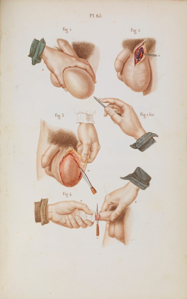
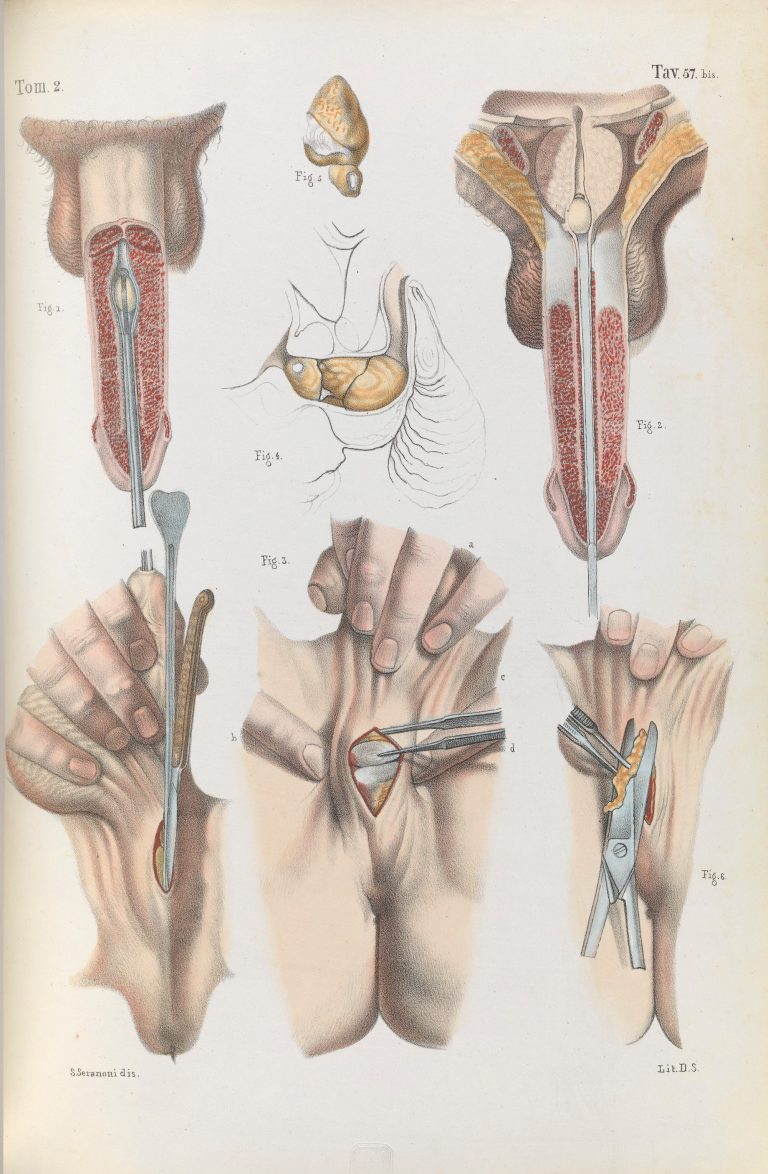
Plate 65, Surgical removal of tumours from the scrotum.
Many of the colour lithographs below were created for US surgeon Joseph Pancoast’s (November 23, 1805 – March 6, 1882) 1844 book A Treatise on Operative Surgery. The blurb tells us: “A treatise on operative surgery comprising a description of the various processes of the art, including all the new operations; exhibiting the state of surgical science in is present advanced condition; with eighty plates containing four-hundred and eighty-six separate illustrations.” These images are for the book’s second edition of 1846, for which they were “enlarged”. Other images can be found in the 1848 work Précis iconographique de médecine opératoire et d’anatomie chirurgicale by Claude Bernard (1813-1878). They are captivating and unsettling.
Plate LXVI. Surgical technique for lithotomy.
Plate XLIX. Surgery to correct strabismus, involving the division of the internal rectus of the right eye. Strabismus is the misalignment of the eyes.
Plate XXXIV. Surgical instruments used for external urethrotomy in prostatectomy (removal of part of the prostate gland).
Removing the placenta and umbilical cord after birth. Abbildungen aus dem Gesammtgebiete der theoretisch-praktischen Geburtshülfe, nebst beschreibender Erklärung derselben / Nach dem Französischen des Maygrier bearbeitet und mit Anmerkungen versehen von Eduard Casp. Jac. von Siebold – 1829
Plate XLVIII. Illustration of surgery on the eye for the removal of a cataract. Operation by extraction – inferior section of the cornea.
Plate LVI. Surgery for the removal of the mammary gland. 19th Century. Iconografia d’anatomia chirurgica e di medicina operatoria / [Jean Baptiste Marc Bourgery]
Plate 42, Techniques for the removal of cataracts.
Plate 70, Surgical techniques for lithotripsy (the removal of bladder and kidney stones). Précis iconographique de médecine opératoire et d’anatomie chirurgicale by Claude Bernard (1848).
Plate LXVII. Surgical technique for lithotomy (the removal of a bladder stone). Bilateral and vesico-rectal operation.
Fig. 1. Removal with the forceps by torsion and traction. Fig. 2, 3. Removal by ligature. Fig. 4,5. Simple hare-lip. Fig. 6, 7, 8. ‘Double hare-lip’ and ‘complicated hare-lip’.
Plate 65, Surgical removal of tumours from the scrotum.